
Case Study
Porcelain Veneers
The Challenge
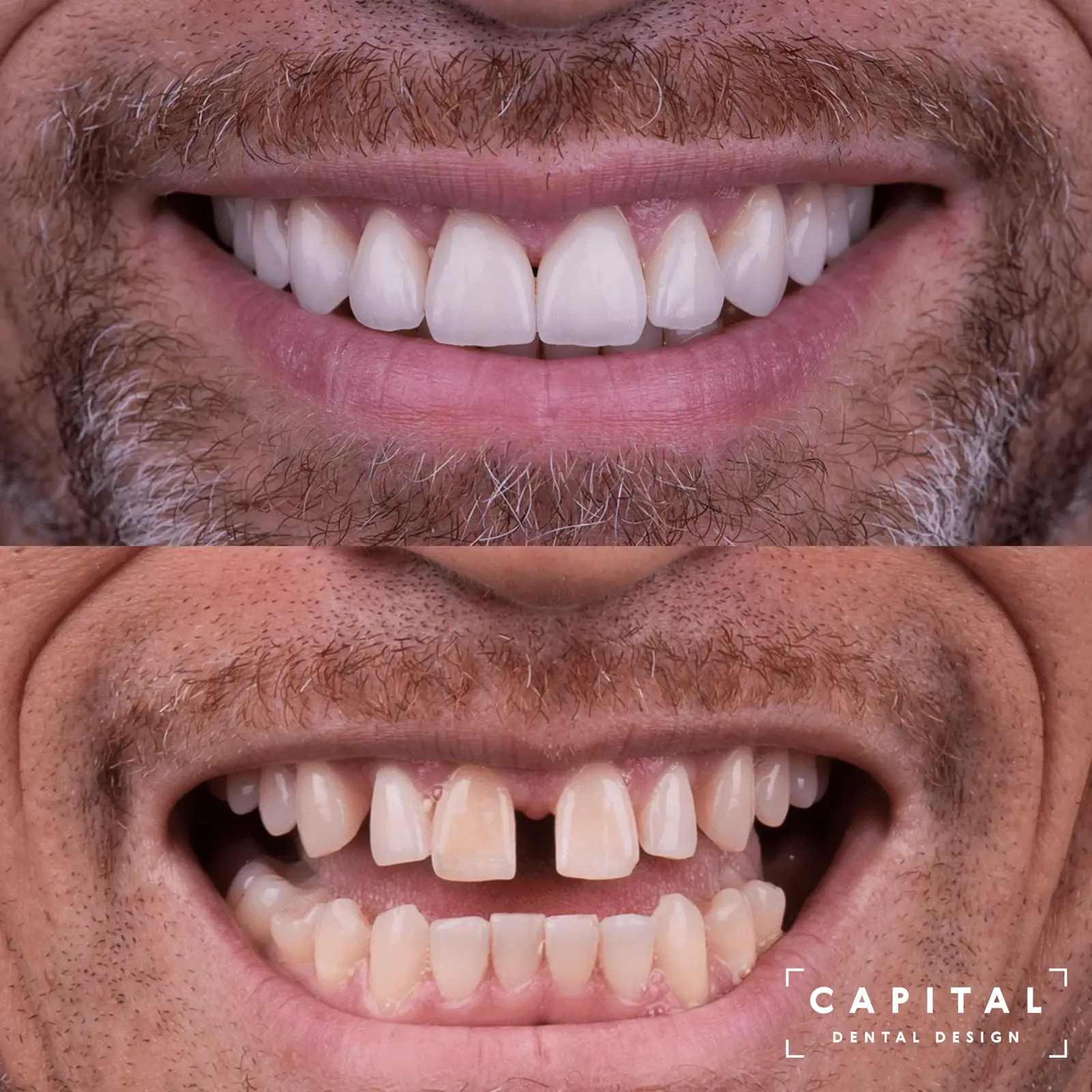
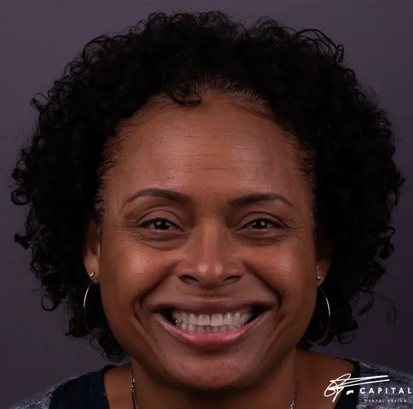
The patient presented with uneven tooth edges and compromised color caused by years of functional wear and nighttime grinding. Despite sound underlying tooth structure, the smile had lost its harmony and vitality. The goal was to restore natural proportions, symmetry, and shade while preserving as much healthy enamel as possible.
12 Steps
From initial presentation through final delivery. Every phase is planned, verified, and documented before proceeding to the next.
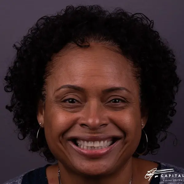
Comprehensive photography and clinical evaluation document the starting point. The patient's primary concerns are mapped and measured. High-resolution imaging captures every detail of tooth shape, color, texture, and gum architecture that will inform the design.
Detailed diagnostic analysis of the bite, tooth proportions, gum symmetry, and facial reference points. Digital measurements establish the mathematical framework for a predictable outcome. Every asymmetry is identified and a correction strategy is mapped.
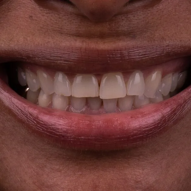

Using Digital Smile Design protocol, the new smile is drawn over the patient's photograph. Tooth length, width, midline position, and smile arc are designed in relation to the face. The patient reviews and approves the design before any clinical work begins.
The macro design is refined at the individual tooth level. Each tooth receives specific proportion adjustments. Surface texture, line angles, and translucency zones are specified for the ceramist. This is the blueprint that guides every subsequent clinical step.
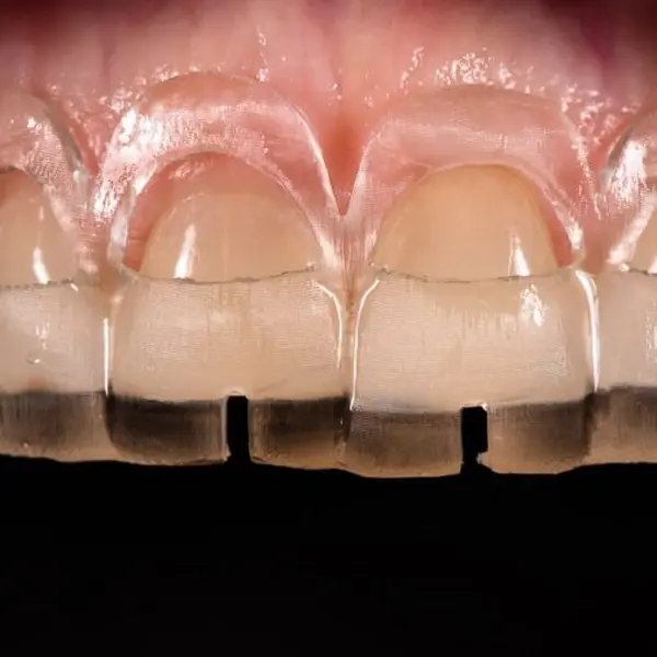
A digitally designed surgical guide ensures gum tissue is recontoured to the exact specifications of the smile design. Crown lengthening creates the correct tooth proportions and symmetrical gum line that will frame the final veneers. Precision here is measured in tenths of a millimeter.
Immediately following gum surgery, the tissue position is verified against the design plan. A healing period allows the tissue to mature and stabilize in its new position. Only after complete healing do we proceed with veneer preparation, ensuring the final framework is stable.
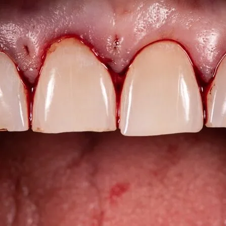
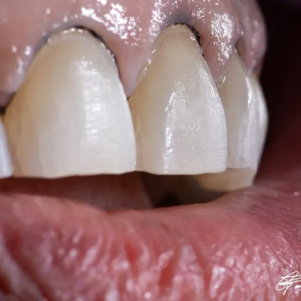
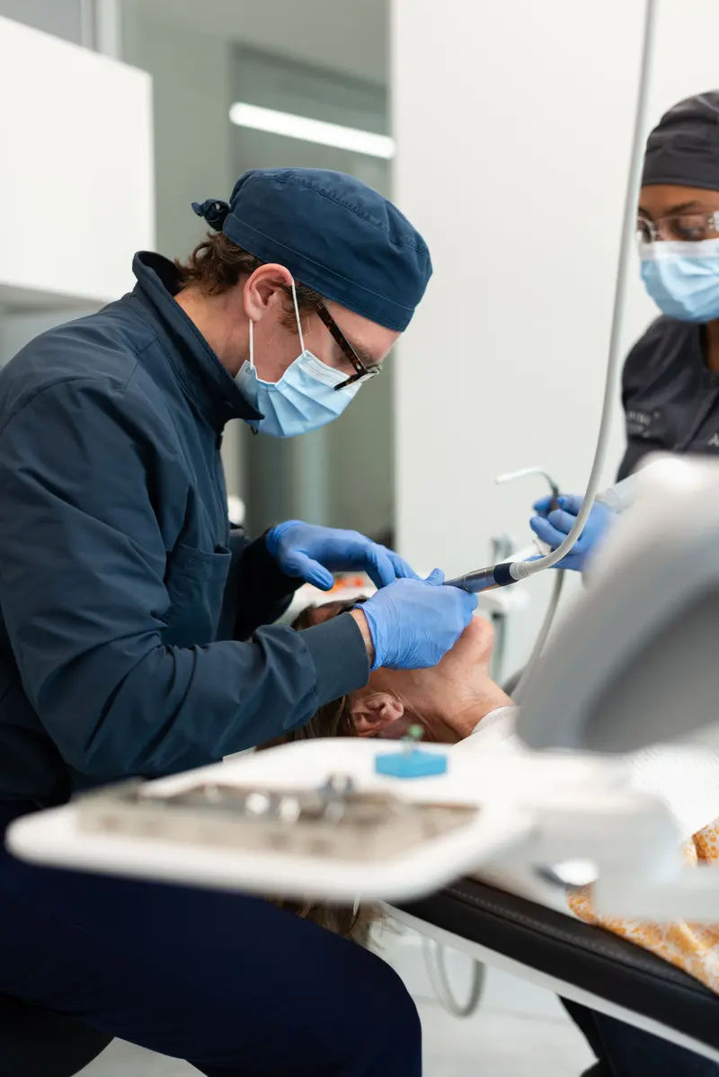
Under 25x microscope magnification, each tooth is prepared with just 0.3mm of reduction. This minimally invasive approach preserves maximum enamel while creating the precise space needed for ultra-thin porcelain. Every preparation margin is verified under the microscope for absolute accuracy.
Total isolation with rubber dam ensures a completely dry field for bonding. Each veneer is individually etched, silanated, and bonded with light-cured resin cement under controlled conditions. The bonding protocol is critical to long-term durability and creates a molecular bond between porcelain and enamel.
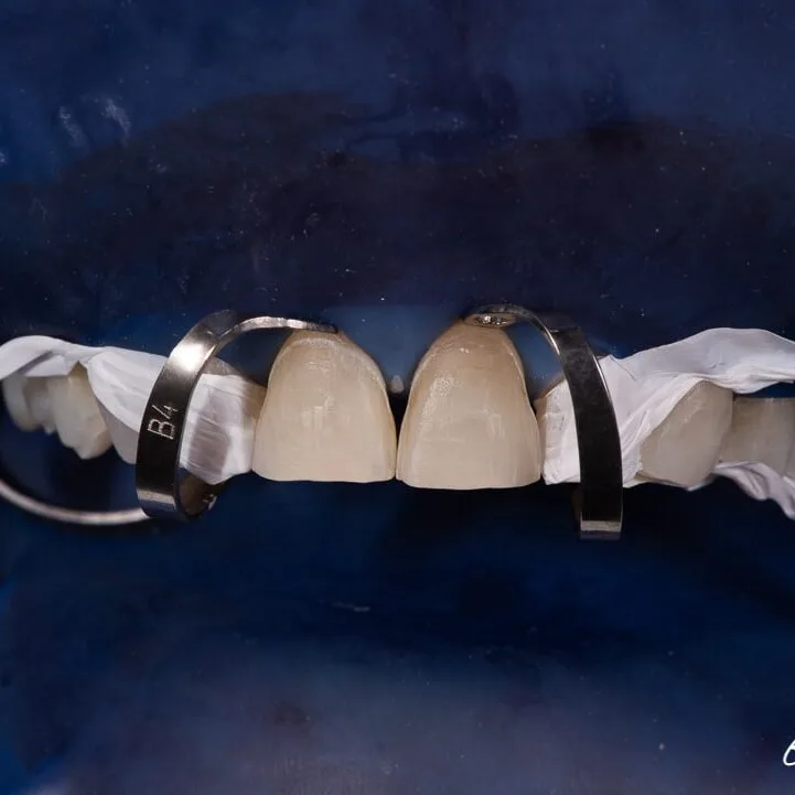
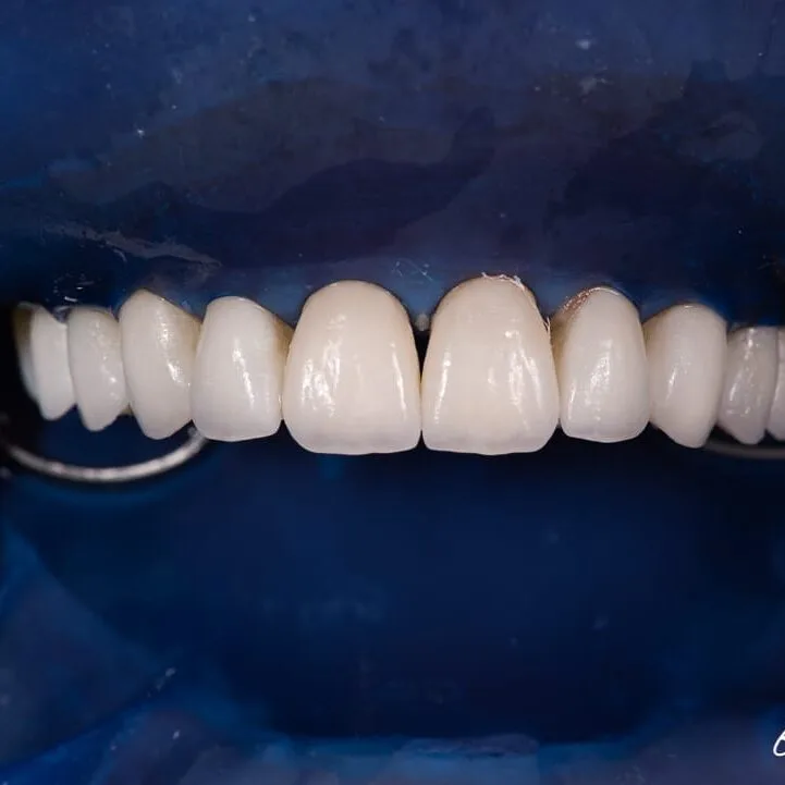
Isolation is removed and the veneers are polished and refined. Margins are verified, contacts are adjusted, and the bite is checked in all excursive movements. The immediate result reveals the transformation from worn and uneven to harmonious and natural.
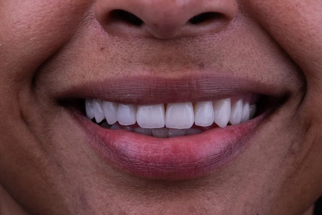
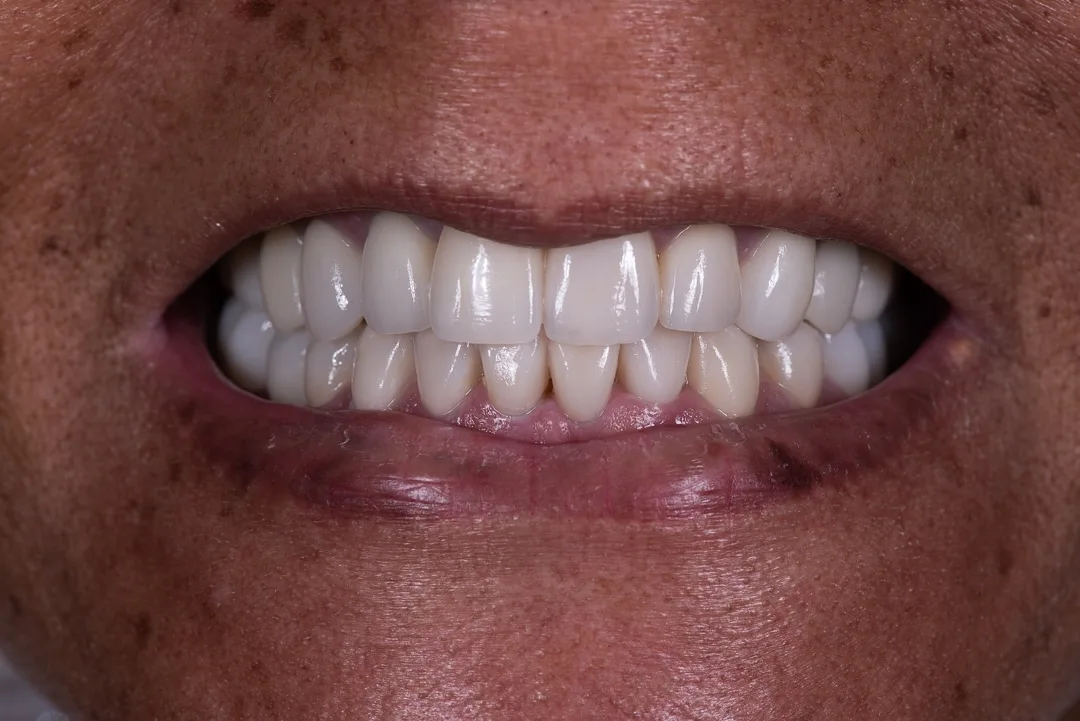
Close-up photography at high magnification evaluates surface texture, shade transitions, and translucency. The porcelain mimics the natural micro-anatomy of teeth with surface ridges, perikymata lines, and color gradients that make the veneers indistinguishable from natural dentition.
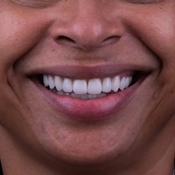

The complete smile is evaluated against the original design plan. Tooth proportions, gum symmetry, midline alignment, and smile arc are verified. The final result matches the approved Digital Smile Design, confirming that the planning process produced a predictable and harmonious outcome.
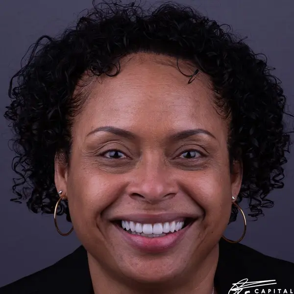
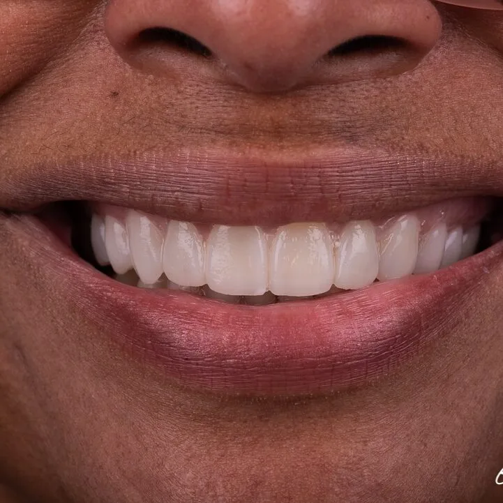
The completed case photographed in natural light. A confident, natural smile that integrates with the patient's face and features. Minimally prepared porcelain veneers, bonded under total isolation, designed digitally before a single tooth was touched. This is design-driven dentistry.
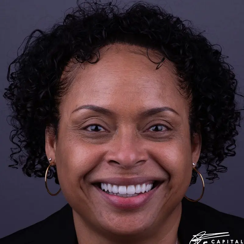
The Transformation
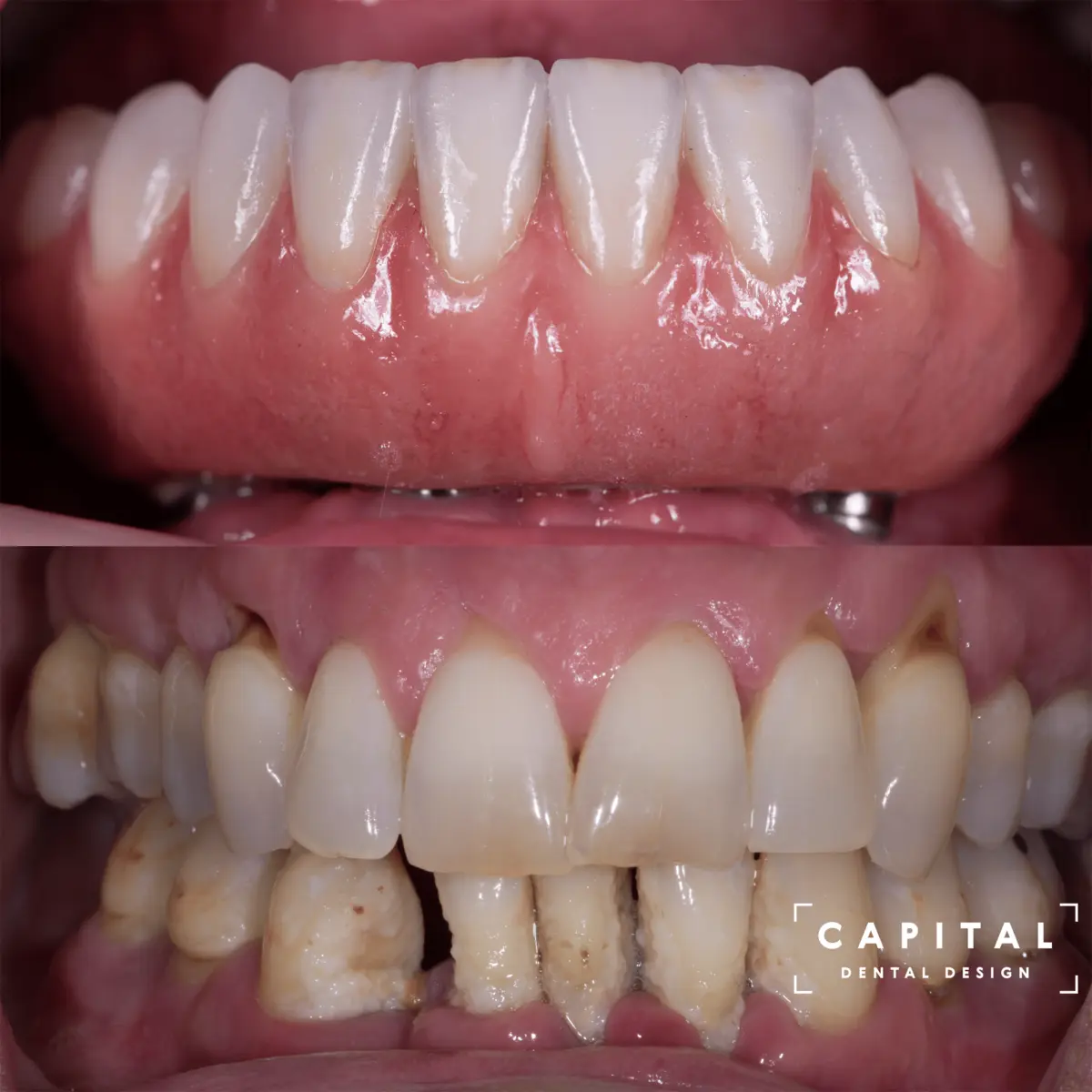
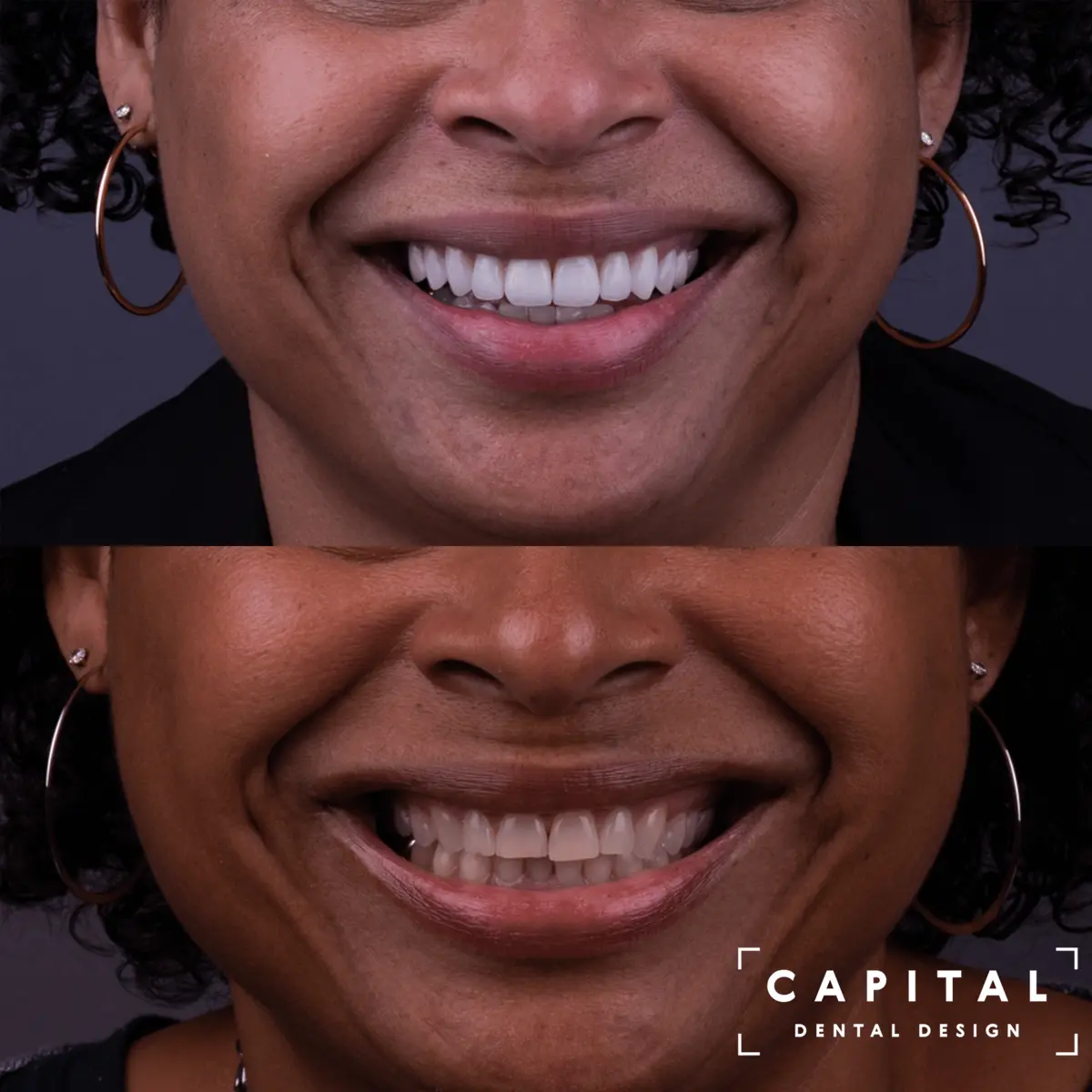
Side by side comparison reveals the scope of the transformation while maintaining the natural character of the smile.
Before
After

Address
6946 Forest Ave, Suite 205
Richmond, VA 23230
Phone
804.320.8894Hours
Monday – Thursday
9:00 AM – 5:00 PM